Recombinant Human CD2 protein ,C- His Tag
  • 1
ATMP00031HU
-
+
Spot
Inquiry
  • Product Details
  • Frontier progress
  • How to order
  • Instructions
Summaryicon
English name
Recombinant Human CD2 protein ,C- His Tag
Purity
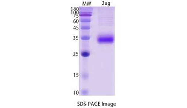
>90% as determined by SDS-PAGE
Endotoxin level
<1.0 EU per μg of the protein as determined by the LAL method.
Construction
A DNA sequence encoding the human CD2(Met1~Asp209) was fused with the C-terminal His Tag
Accession #
P06729
Host
Mammalian cells
Species
Homo sapiens (Human)
Predicted Molecular Mass
24.73KDa
Formulation
Supplied as solution form in PBS, pH7.5 or lyophilized from PBS, pH7.5.
Shipping
In general, proteins are provided as lyophilized powder/frozen liquid. They are shipped out with dry ice/blue ice unless customers require otherwise.
Stability &Storage
Use a manual defrost freezer and avoid repeated freeze thaw cycles.
Store at 2 to 8 °C for one week .
Store at -20 to -80 °C for twelve months from the date of receipt.
Reconstitution
Reconstitute in sterile water for a stock solution.A copy of datasheet will be provided with the products, please refer to it for details.
SDS-PAGE imageicon
    Backgroundicon
    Background
    T-cell surface antigen CD2 is also known as Erythrocyte receptor, LFA-2, LFA-3 receptor, Rosette receptor, T-cell surface antigen T11/Leu-5 and SRBC, is a single-pass type I membrane protein found on the surface of T cells and natural killer (NK) cells. CD2 is a member of the immunoglobulin superfamily. CD2 / SRBC contains 1 Ig-like C2-type (immunoglobulin-like) domain and 1 Ig-like V-type (immunoglobulin-like) domain. CD2 / SRBC interacts with other adhesion molecules, such as lymphocyte function-associated antigen-3 (LFA-3 / CD58) in humans, or CD48 in rodents, which are expressed on the surfaces of other cells. In addition to its adhesive properties, CD2 also acts as a co-stimulatory molecule on T and NK cells. CD2 is a specific marker for T cells and NK cells, and can therefore be used in immunohistochemistry to identify the presence of such cells in tissue sections.
    Alternative Names
    CD2,SRBC,LFA-2,T11
    References
    Sridharan, Seawright, Landes, Cao, Singh, Davis, Mao, Singh, Zhang, Nelson, Boerma (2020) Effects of single-dose protons or oxygen ions on function and structure of the cardiovascular system in male Long Evans rats Life sciences in space research 26() 62-68
    Studies are required to determine whether exposures to radiation encountered during manned missions in deep space may have adverse effects on the cardiovascular system. Most of the prior studies on effects of simulated space radiation on the heart and vasculature have been performed in mouse models. To provide data from a second animal species, two studies were performed to assess effects of high-energy charged particle radiation on the heart and abdominal aorta in a rat model. In study A, male Long Evans rats were exposed to whole-body protons (250 MeV, 0.5 Gy) or oxygen ions (16O, 600 MeV/n, 0.5 Gy), and ultrasonography was used to measure in vivo cardiac function and blood flow parameters at 3, 5, 9 and 12 months after radiation, followed by tissue collection at 12 months. In study B, male Long Evans rats were exposed to 16O (1 GeV/n, 0.01-0.25 Gy), and hearts collected at 6 to 7 and 12 months for histology and western-blots. Both protons (250 MeV) and 16O (600 MeV/n) caused a decrease in left ventricular posterior wall thickness at 3-5 months, but did not change echocardiographic measures of cardiac function. In Pulsed-wave Doppler assessment of the abdominal aorta, an increase was seen in mean velocity, peak velocity, and velocity time integral at 12 months after 16O (600 MeV/n), suggesting a change in vascular function. There were no significant changes in histopathology or histological quantification of total collagens in heart or aorta. On the other hand, an increase was seen in a 75 kDa peptide of collagen type III in the left ventricle of rats exposed to protons (250 MeV) and 16O (600 MeV/n and 1 GeV/n), suggesting that radiation caused remodeling of existing collagens in the heart. 16O (600 MeV/n and 1 GeV/n) caused increases in left ventricular protein levels of immune cell markers CD2, CD4, CD8, and CD68. A single low dose of whole body protons or 16O in male Long Evans rats did not change cardiac function or induce gross pathological changes in the heart or aorta, but induced mild changes in vascular function and remodeling of existing collagens in the heart. Altogether, studies in prior mouse models and the current work in rats indicate minor changes in cardiac function and structure after a low dose of single-ion radiation.

    Guarantee and Disclaimer:

    After receiving the product, if finds that the product is mismatched, damaged or missing components, please keep the original package and submit the objection to the company by mail within seven working days. Failure to file an objection within the time limit is considered qualified.

    When the buyer keeps the product, it should be kept in accordance with the storage conditions shown in the product label and the manual. If the product quality is caused by improper storage, it will not be guaranteed.

    When the buyer tests the product, they should be submitted by the beginning of use when found the quality issue, rather than when the product was used or used up, in order to prepare our products for recycling and confirm the quality of the products. If it is the quality problem, our company is responsible for exchange or return. In the event of a claim, our company will compensate the discretion within the scope of the product price limit and will not accept any part of the value of the product itself. Since the date of receipt, the product has not been reflected for more than three months and should not be returned.

    The products provided by our company are for research use only and should not be used for clinical diagnosis or treatment. If a unit or individual changes the use of our products without authorization, we will not bear any responsibility.