AtaGenix

Recombinant Human PDCD1 protein ,C- His Tag
  • 1
ATMP00045HU
-
+
Spot
Inquiry
  • Product Details
  • Frontier progress
  • How to order
  • Instructions
Summaryicon
English name
Recombinant Human PDCD1 protein ,C- His Tag
Purity
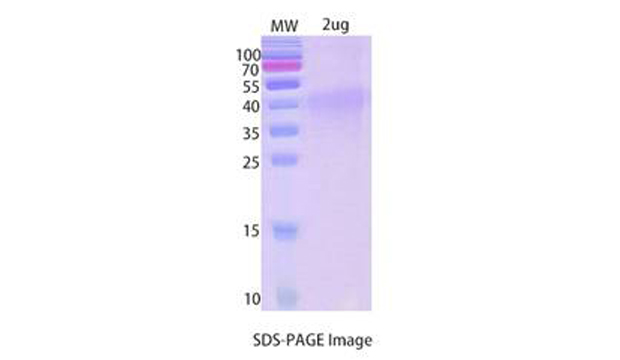
>90% as determined by SDS-PAGE
Endotoxin level
<1.0 EU per μg of the protein as determined by the LAL method.
Construction
A DNA sequence encoding the human PDCD1(Met1~Val170) was fused with the C-terminal His Tag
Accession #
Q15116
Host
Mammalian cells
Species
Homo sapiens (Human)
Predicted Molecular Mass
20.04kDa
Formulation
Supplied as solution form in 150 mM NaCl, 50 mM Tris-HCl, pH 7.5 or lyophilized from 150 mM NaCl, 50 mM Tris-HCl, pH 7.5.
Shipping
In general, proteins are provided as lyophilized powder/frozen liquid. They are shipped out with dry ice/blue ice unless customers require otherwise.
Stability &Storage
Use a manual defrost freezer and avoid repeated freeze thaw cycles.
Store at 2 to 8 °C for one week .
Store at -20 to -80 °C for twelve months from the date of receipt.
Reconstitution
Reconstitute in sterile water for a stock solution.A copy of datasheet will be provided with the products, please refer to it for details.
SDS-PAGE imageicon
    Backgroundicon
    Background
    Programmed cell death protein 1 (PD-1) is also known as CD279 and PDCD1, is a type I membrane protein and is a member of the extended CD28/CTLA-4 family of T cell regulators. PDCD1 is expressed on the surface of activated T cells, B cells, macrophages, myeloid cells and a subset of thymocytes. PD-1 has two ligands, PD-L1 and PD-L2, which are members of the B7 family. PD-L1 is expressed on almost all murine tumor cell lines, including PA1 myeloma, P815 mastocytoma, and B16 melanoma upon treatment with IFN-γ. PD-L2 expression is more restricted and is expressed mainly by DCs and a few tumor lines. PD1 inhibits the T-cell proliferation and production of related cytokines including IL-1, IL-4, IL-10 and IFN-γ by suppressing the activation and transduction of PI3K/AKT pathway. In addition, coligation of PD1 inhibits BCR-mediating signal by dephosphorylating key signal transducer. In vitro, treatment of anti-CD3 stimulated T cells with PD-L1-Ig results in reduced T cell proliferation and IFN-γ secretion. Monoclonal antibodies targeting PD-1 that boost the immune system are being developed for the treatment of cancer.
    Alternative Names
    PDCD1,PD1,CD279,SLEB2
    References
    Tian, Yang, Han, He, Liao (2020) A novel immune checkpoint-related seven-gene signature for predicting prognosis and immunotherapy response in melanoma International immunopharmacology 87() 106821
    New emergence of immunotherapy has significantly improved clinical outcome of melanoma patients with advanced and metastatic diseases. We aimed to develop a gene signature based on the expression of PD-1/PD-L1 signaling pathway genes to predict prognosis and immunotherapy response in melanoma patients. Melanoma samples from The Cancer Genome Atlas (TCGA) database and Gene Expression Omnibus (GEO) were used as the training set and external validation sets respectively. Prognostic genes for overall survival (OS) were identified by univariate Cox regression analysis. Then a multi-gene risk signature was established with the Least Absolute Shrinkage and Selector Operation (LASSO) regression and multivariate Cox regression. The predictive and prognostic value of gene signature was evaluated by Kaplan Meier curve, Time-dependent receiver operating characteristic (ROC) curve, and area under curve (AUC). Gene set enrichment analysis (GSEA) was performed to investigate the discrepantly enriched biological processes between low-risk and high-risk group of melanoma patients. A seven-gene risk signature (BATF2, CTLA4, EGFR, HLA-DQB1, IKBKG, PIK3R2, PPP3CA) was constructed. The signature was an independent risk factor for OS (hazard ratio = 1.544, p <0.001) and it could robustly predict OS in both training and validation sets. Besides, high risk scores indicated advanced clinical stage and no response to immunotherapy for melanoma patients. GSEA demonstrated that high risk score was intimately associated with immune response and immune regulation. In conclusion, the novel seven-gene signature could serve as a robust biomarker for prognosis and a potential indicator of immunotherapy response in melanoma.

    Guarantee and Disclaimer:

    After receiving the product, if finds that the product is mismatched, damaged or missing components, please keep the original package and submit the objection to the company by mail within seven working days. Failure to file an objection within the time limit is considered qualified.

    When the buyer keeps the product, it should be kept in accordance with the storage conditions shown in the product label and the manual. If the product quality is caused by improper storage, it will not be guaranteed.

    When the buyer tests the product, they should be submitted by the beginning of use when found the quality issue, rather than when the product was used or used up, in order to prepare our products for recycling and confirm the quality of the products. If it is the quality problem, our company is responsible for exchange or return. In the event of a claim, our company will compensate the discretion within the scope of the product price limit and will not accept any part of the value of the product itself. Since the date of receipt, the product has not been reflected for more than three months and should not be returned.

    The products provided by our company are for research use only and should not be used for clinical diagnosis or treatment. If a unit or individual changes the use of our products without authorization, we will not bear any responsibility.